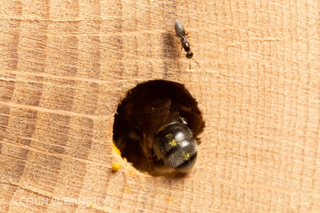
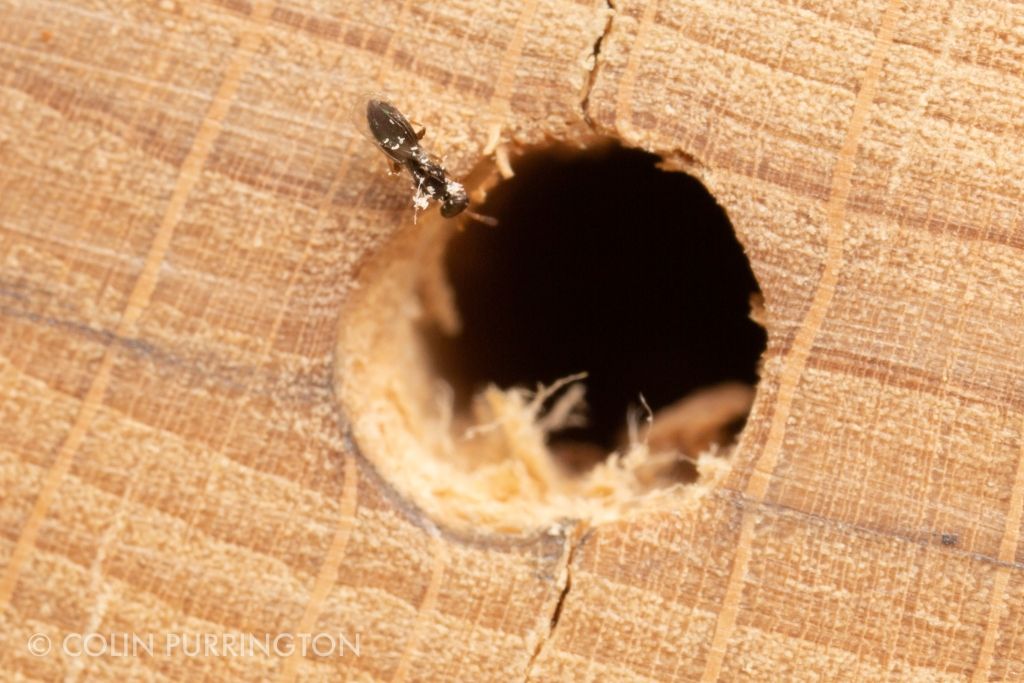
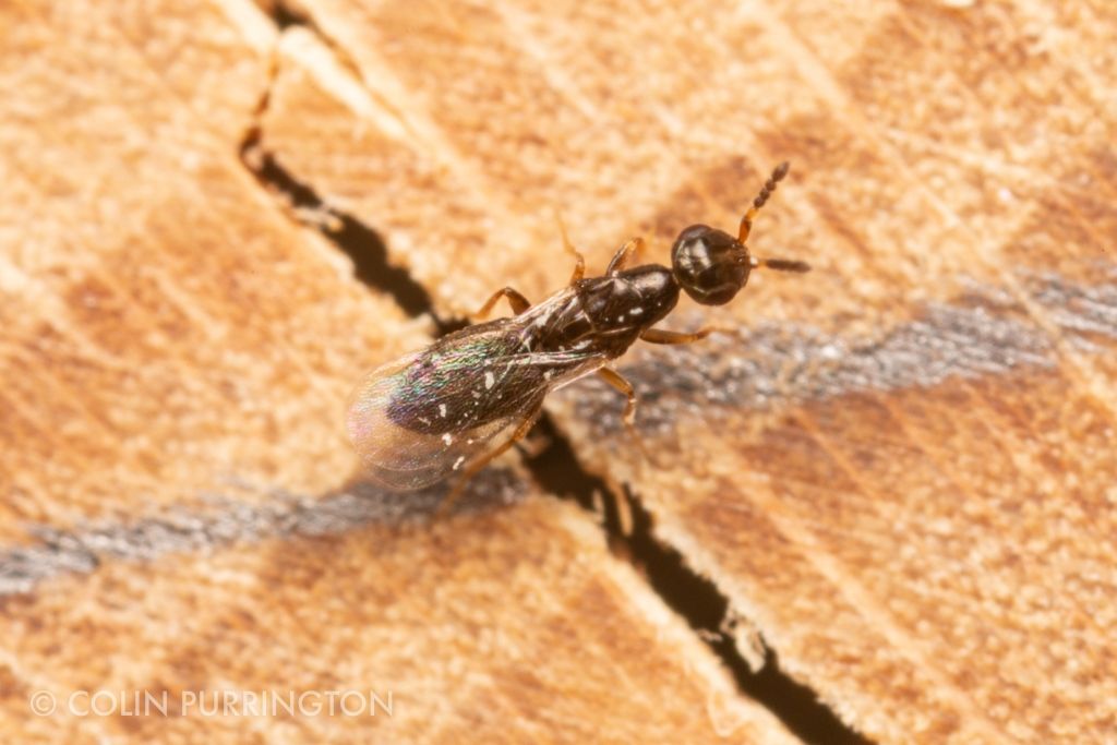
I recently set up a mason bee house, and almost instantly attracted a parasitoid, a female Melittobia. She was so tiny that I first thought it was a thrips. But its behavior was odd and definitely not thripsy. The insect would linger near the holes that were being used by mason bees (Chelostoma philadelphi and Osmia sp.), approach, then scurry away, then repeat. On one occasion it peered down an unoccupied hole and paused for several seconds before walking away. It was covered in wood shavings, too, so I inferred that she’d been inside the holes on at least one occasion. All this activity screamed, “parasitoid”, of course, so I had to get some pictures:


 I’m not sure which species she is (there are 14 according to González et al. 2004), but I gather that all members of the genus can parasitize mason bees. Females spend up to 48 hours lurking around a hole (“assessing” per González article) and then puncture a developing larvae with her ovipositor, drink some of the hemolymph, and deposit eggs. There’s a pause in between the hemolymph-feeding and the egg-laying, presumably to allow some of the nutrients to be allocated to eggs. Females are not at all picky about host larvae. Recorded hosts for M. acasta include honeybees, bumblebees, leafcutting bees, and various wasps, flies, beetles, and butterflies (González et al. 2004).
Things get exciting when the wasps eclose, with the early-emerging male killing the slackers that are still developing. There are two types of females: brachytperous (reduced wings) and macroptyrous (regular wing size). I’m assuming the one(s) I viewed are the regular kind.
I’ll post more photographs if I can get them, but whatever is going on inside the holes is of course hidden. But I’m going to make a few glass-sided mason bee houses in the next few weeks so hopefully I can get some larval pics one of these years. This spotting is a reminder to use paper tube inserts whenever possible so that larvae can be examined at the end of the season for parasites. I’m rather interested in parasites but I’d like to encourage a healthy mason bee population when possible.
Much thanks to Ross Hill for identification, and of course to people behind BugGuide for facilitating such IDs. These observations are also logged into iNaturalist.


