Here are photographs of my insect hotel from Spring 2022. It has three levels and features nesting holes of varying diameters so that I can attract multiple species of bees and wasps. If you’d like to see what shows up this spring and summer, follow me on iNaturalist — I post photographs of everything that arrives, plus post photographs of what is inside the nesting tubes at the end of season. The latter is possible because all holes are lined with paper straws or are stems that can be split open. Having nests that can be dismantled also allows me to minimize the number of unwanted parasites and diseases that might otherwise thrive.
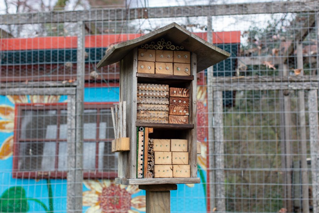
Starting from the top, in the attic space, are 7″ sections cut from a princess tree (Paulownia tomentosa) growing in my yard. The wood blocks have slightly smaller-diameter holes (1/2″) and are lined with 6″ lengths of paper bubble-tea straws. Together, these holes will likely attract grass-carrying wasps (Isodontia spp.), eastern carpenter bees (Xylocopa virginica), and sculptured resin bees (Megachile sculpturalis). And, probably, jumping spiders.
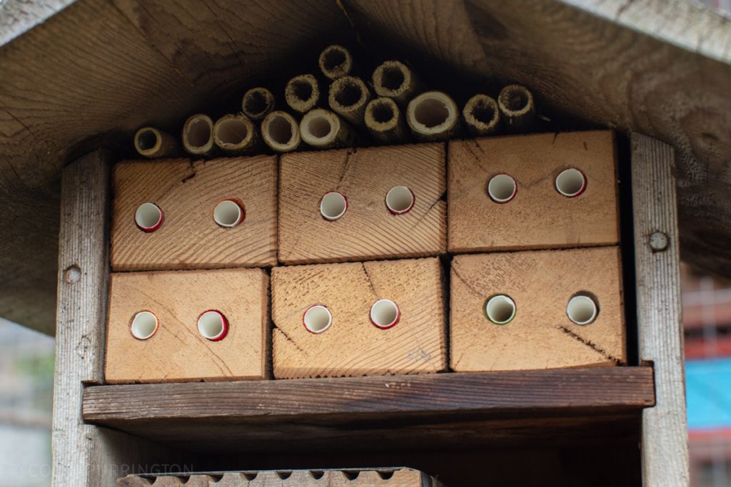
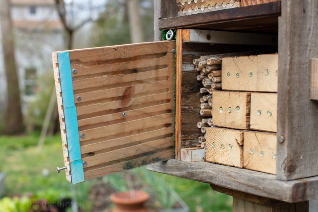
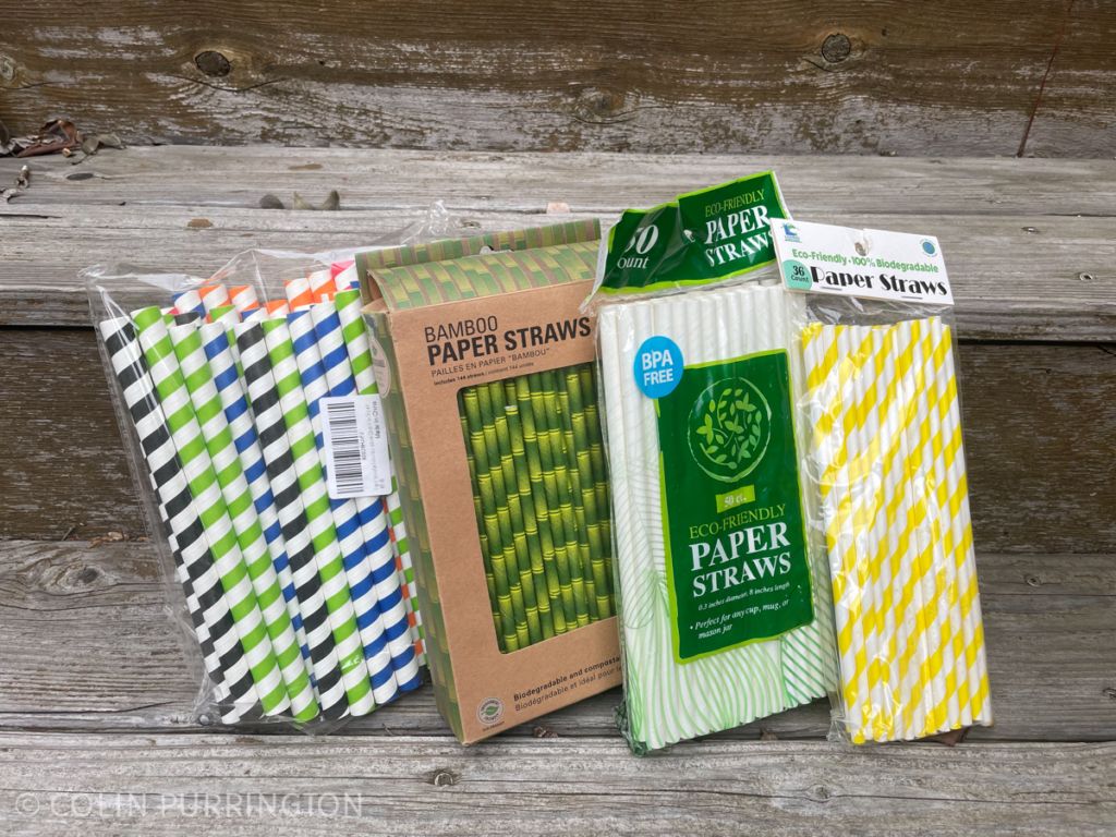
The middle section has more traditional offerings, with paper tubes inside nesting trays (I bought them online) and inside blocks with 6″ holes that I drilled. You can buy super-hard paper liners made for insect hotels, or you can be cheap like me and just use paper drinking straws. I found straws online that are a tad shy of 5/16″. There are dozens of bee and wasp species that will use holes of this size. Note that if you splurge on the trays, you’ll need a way to compress them; I use pipe clamps but luggage straps work, too.
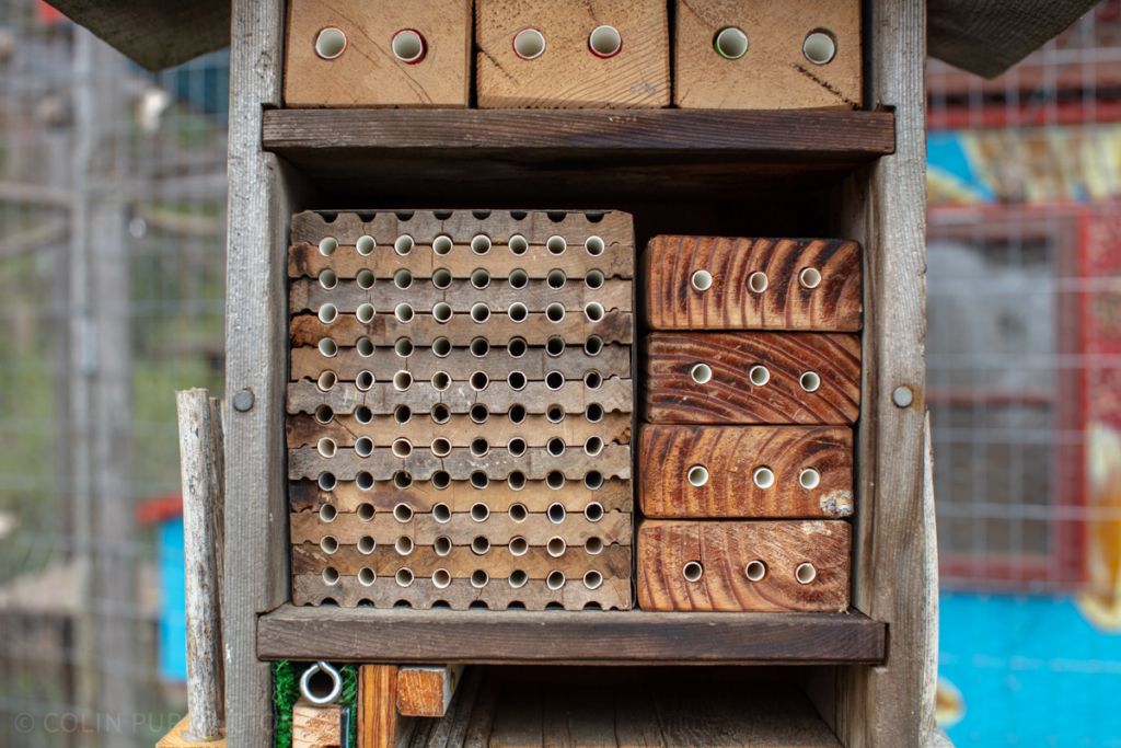
The lower level has cavities that are slightly smaller still, to attract bees and wasps that have smaller body sizes. The blocks have 1/4″ holes fitted with paper drinking straws that are just shy of that. Stems to the left of the blocks are 7″ sections of swamp milkweed (Asclepias incarnata) from my garden (I also like bee balm). These smaller nests seem to attract Georgia mason bees (Osmia georgica) and my favorite wasp, Trypoxylon collinum (the male stays home to guard the nest).
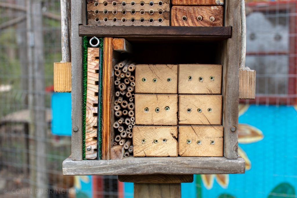
Also on the lower level are square holes routered onto a pine board and covered with Plexiglas. This allows me to spy on the bees and wasps as they construct and provision their nests. It’s early in the season so there’s no real excitement in the photograph, but you can see horned-faced mason bees (Osmia cornifrons) in tunnels #2 and #8. They are both males and are waiting for the females to show up. This panel can be taken apart at the end of the season and cleaned.
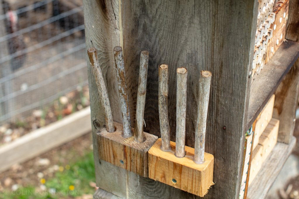
Finally, I have an array of stems on both sides of the insect hotel to accommodate species that might prefer to nest vertically. I’m hoping to attract some leafcutting bees (Megachile), but they are said to dislike the commotion at large insect hotels. One can always dream.
Using paper straws to line nests
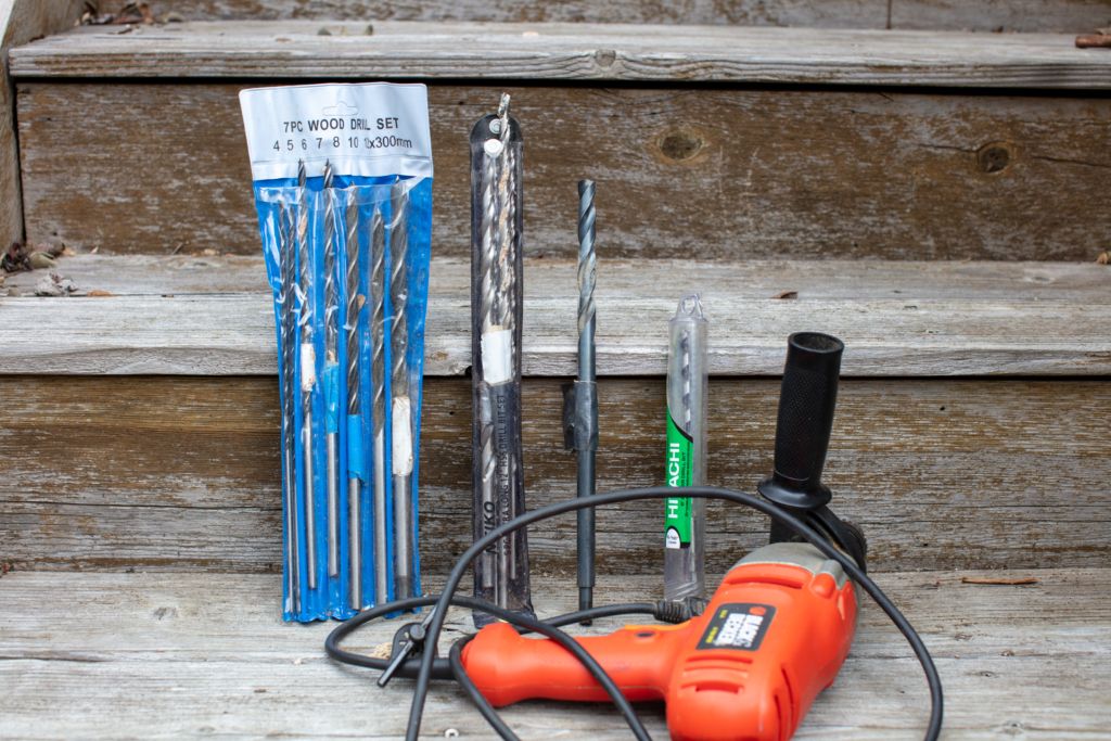
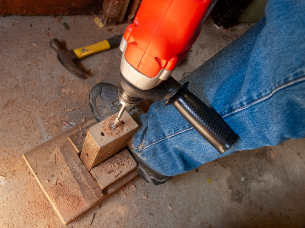
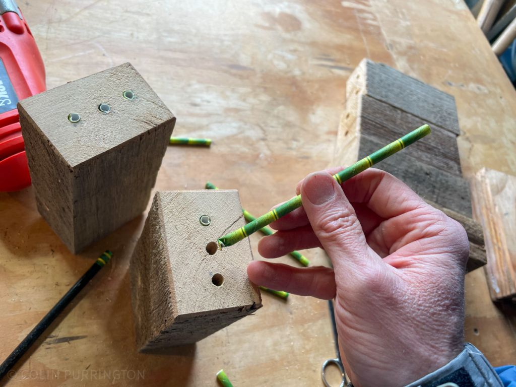
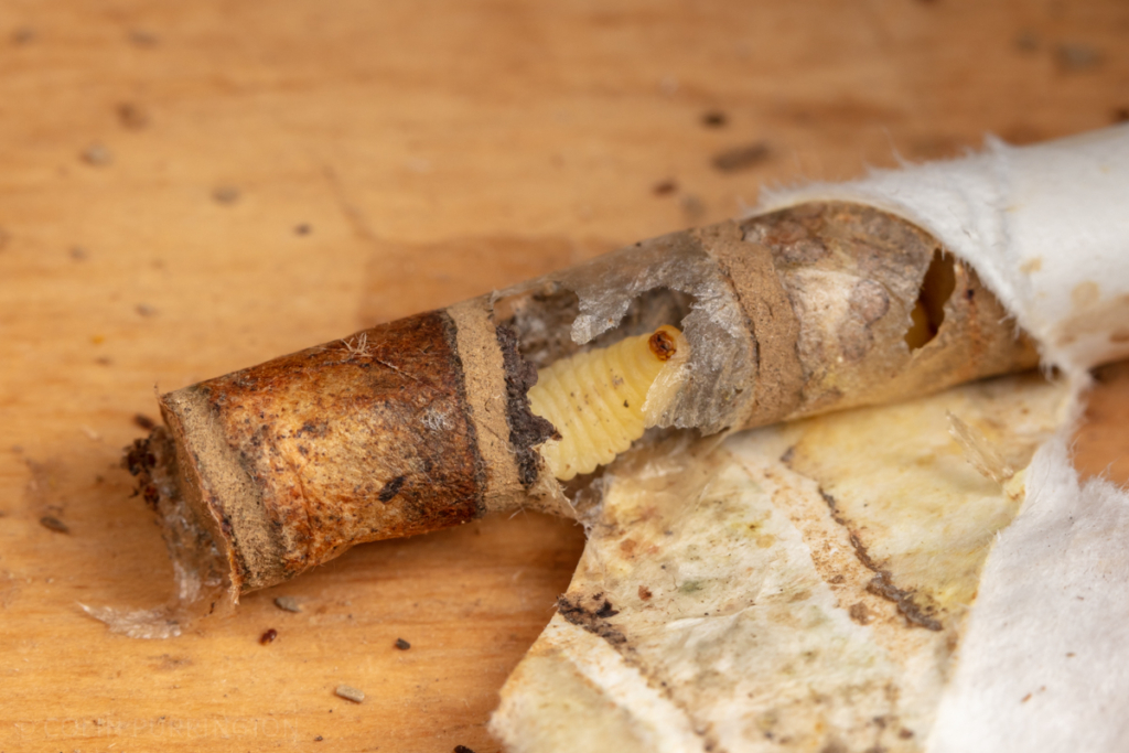
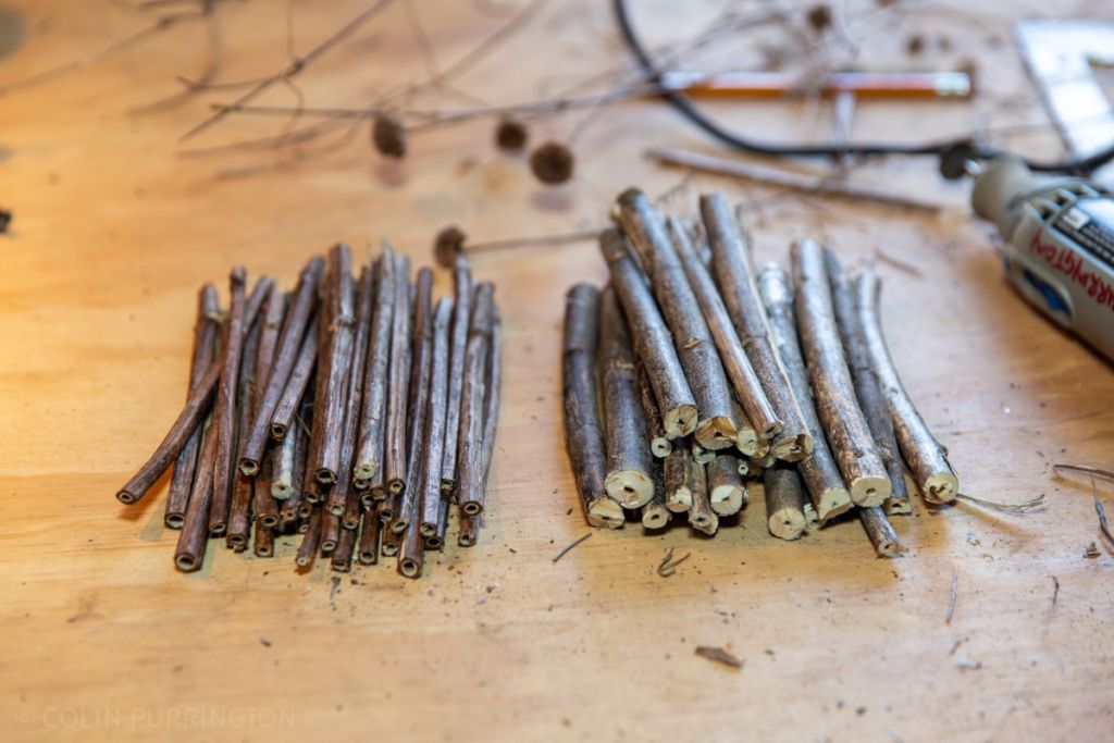
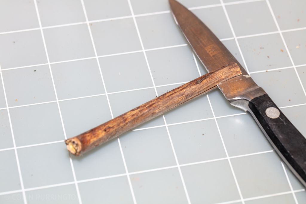
If you want to make your own insect hotel you’ll need to invest in some long drill bits so that your tunnels can be at least 6″ long. I purchased both a standard set (inches) and a metric set, plus two individual bits that I purchased separately. And you should make yourself a little floor brace so that you can drill without the block rotating wildly. For straws, there are dozens of online places to get paper ones, but I’ve also found them at local grocery stores. And when you get to the stage of inserting the straws into blocks, trim the lengths and then crimp the end (see pic, below) so that the inhabitants don’t extend their nesting materials into the block itself; this makes it easier to use a forceps to pull the straw out. Then during the winter, unroll the straw to reveal contents. Save the pupae you want (in small containers, outside, so they develop normally) and discard parasites such as Houdini flies.
Using stems
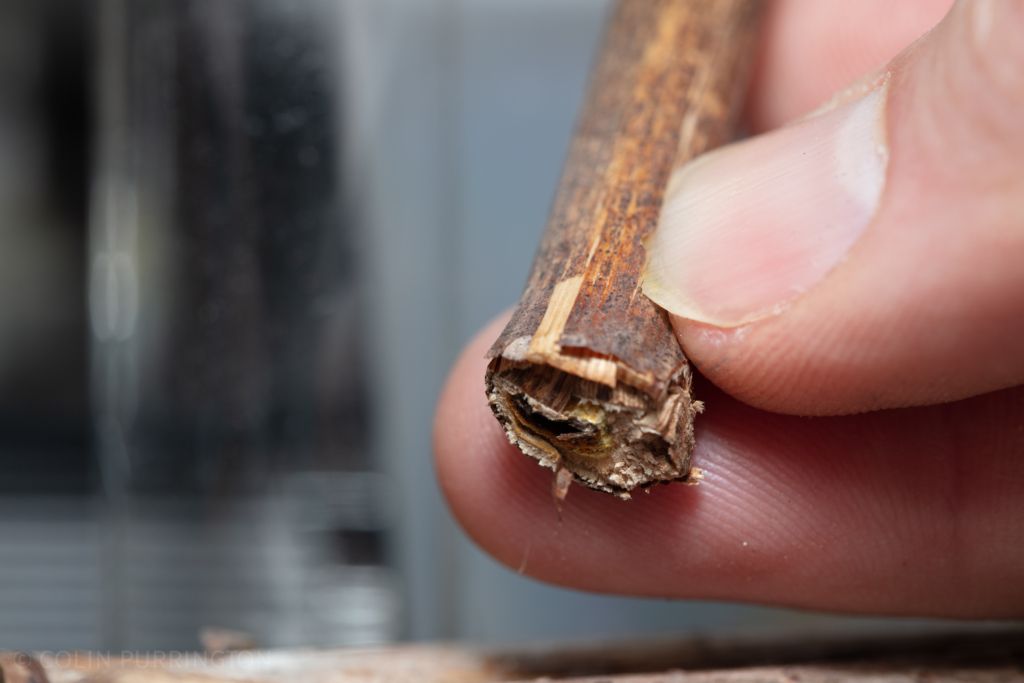
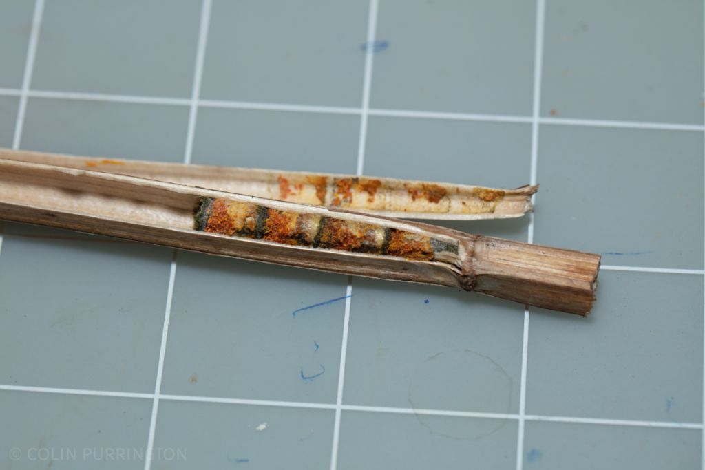
For making reeds, I highly recommend using a rotary tool with a cut off blade instead of pruners or saws, both of which leave jagged edges and sometimes cause fractures that can allow parasites to get inside the stem. When you’re ready to sort through the stems, use a knife to split open the stem, then use your thumbnail to continue the break to the end. Contents can be cleaned and sorted just as for those inside straws.
Identifying contents
Part of the fun of having an insect hotel is it provides a nice window to all the organisms you might not even know are in your yard. I’ve managed hotels for over a decade and there is always something new each year, and during a pandemic these surprises keep me going. E.g., I found a parasitic beetle last year. So I highly recommend taking photographs of your hotel guests and uploading them onto iNaturalist (free), a site that can often get your organisms identified by other like-minded folks. And I always recommend buying some books. Here are the titles I own:
- Wasps: A Guide for Eastern North America (Heath Holm)
- Bees: An Identification and Native Plant Forage Guide (Heather Holm)
- Common Native Bees of the Eastern United States (Heather Holm)
- The Bees in Your Backyard (Joseph S. Wilson & Olivia Messinger Carril)